Capabilities
- High spatial resolution: The LLSM can achieve higher resolution than a standard light-sheet microscope due to the reduced thickness of the lattice light-sheet beam. Our microscope can achieve up to 230 nm x 230 nm x 370 nm resolution in weakly scattering samples. The LLSM also offers a super-resolution (SIM) mode for even higher resolution.
- High temporal resolution: The LLSM can acquire a full 3D image stack in less than a second (depending on the signal to noise ratio). This makes the LLSM particularly well suited for dynamical studies.
- Reduced toxicity/bleaching: Compared with confocal and epi fluorescence widefield microscopes, the LLSM can dramatically reduce photobleaching for living biological samples. The LLSM only illuminates the focal plane of the detection objective, thus reducing photobleaching of fluorophores outside the focal plane.
- Imaging volume: Due to the positions of illumination objective and detective objective, the image field of view can exceed 100 µm x 100 µm. Depending on the sample and required resolution, imaging depth can exceed 50 µm. For large fields of view, a mosaic can be acquired.

Specifications
- Excitation: 7 Laser wavelengths (405/445/488/532/561/590/639 nm)
- Detection: Simultaneous dual-color acquisition
- Depth of field: The length of the lattice light sheet can be varied from 5 µm to about 75 µm. Longer sheets can image deeper, but sacrifice spatial resolution.
- Spatial resolution: For example, with a 488 nm illumination, the resolution can reach 230 nm x 230 nm x 370 nm in standard mode and 150 nm x 230 nm x 280 nm in SIM mode.
Sample preparation
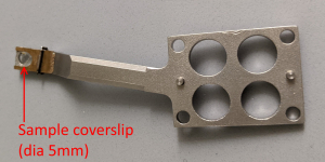
- Sample size: Due to the layout of the illumination and detection objectives, the sample needs to fit on a 5 mm dia glass coverslip and the sample height should not exceed about 2 mm (depends on imaging region and sample orientation).
- Sample must be adhered to the 5 mm dia coverslip.
- The LLSM uses water dipping objectives and images the sample from the top (the water side) without a top coverslip.
- Our LLSM is of the Betzig/Janelia design. For more details on sample prep, please check this link: https://www.aicjanelia.org/post/lattice-light-sheet-sample-prep
Data analysis, visualization, and storage
- Due to the imaging geometry, the raw data from the LLSM is skewed. Hence, the raw data needs to be post-processed for visualization/analysis. Post-processing can be performed on our ALIS workstation, but we can also provide training for processing on your own computer (memory and CPU/GPU requirement can be quite demanding, since datasets can be on the order of 1 TB).
- Because of the size of the datasets, data visualization of LLSM post-processed data is equally challenging. Our workstation is available for simple analysis and visualization with commercial (Imaris, Amira) or open source packages (ImageJ, napari).
- While we provide temporary storage space for processing and visualization, the long-term storage of the acquired data is the responsibility of the user. We will retain raw data for up to 3 months after acquisition or until we reach capacity of our 100 TB storage array (whichever occurs first).
- Transfer of data to your storage location can be manual (bring your own portable drive) or via Globus.
Cost
- LLSM operation: $100.00 fixed setup fee (billed as 2 hr setup time) plus $50.00/hour experiment time. The setup fee is required for each experiment (daily) or after microscope reconfiguration. This fee includes standard post-processing of the data.
- Consulting & Training services: $100.00/hour
- Image analysis: $50.00/hour
Operation
- ALIS staff will align, calibrate, and operate the LLSM.
- Sample mounting is performed by the user (with assistance from staff).
- ALIS staff is available for microscope operation, but the user should be present for imaging guidance. Repeat users can be trained to operate the microscope by themselves.